Second Line of Defence
This level involves cells and chemicals which act against an antigenic structure internally once it has entered the body.
Phagocytosis
Types of Phagocytes
→ Macrophages are mainly involved in the second level of defence, but also have a role in promoting the third level of defence. After they ingest a pathogen they display it's antigen on their surface. This attracts more phagocytes, and also promotes action by T and B cells.
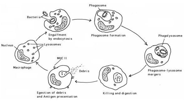
Stages of Phagocytosis
1. The bacterium is attracted to the membrane of the phagocyte.
2. The bacteria are then engulfed by the phagocyte and are encapsulated in a vesicle called a phagosome.
3. Lysosomes form inside the phagocyte and fuse with the phagosome.
4. The enzymes in the lysosome digest the bacteria and break it down.
5. Waste substances are expelled from the cell. Antigenic fragments combine with class 2 MHC markers and are displayed on the surface of the cell.
Phagocytosis
- This process occurs in this context as the removal of all substances and structures which have foreign antigens.
- Phagocytes are different types of white blood cells that are produced in bone marrow.
- This process involves the phagocyte coming in contact with and engulfing the bacterium. Once engulfed inside the phagocyte a vacuole is formed around the bacterium. lysosomes present in the phagocyte fuse with the bacterium and releases digestive enzymes that help to destroy the bacterium.
Types of Phagocytes
- Macrophage
→ Macrophages are mainly involved in the second level of defence, but also have a role in promoting the third level of defence. After they ingest a pathogen they display it's antigen on their surface. This attracts more phagocytes, and also promotes action by T and B cells.
- Neutrophil
- Eosinophil
- Basophil
Stages of Phagocytosis
1. The bacterium is attracted to the membrane of the phagocyte.
2. The bacteria are then engulfed by the phagocyte and are encapsulated in a vesicle called a phagosome.
3. Lysosomes form inside the phagocyte and fuse with the phagosome.
4. The enzymes in the lysosome digest the bacteria and break it down.
5. Waste substances are expelled from the cell. Antigenic fragments combine with class 2 MHC markers and are displayed on the surface of the cell.
Inflammatory Response
Agents Involved
Stages of the Inflammatory Response
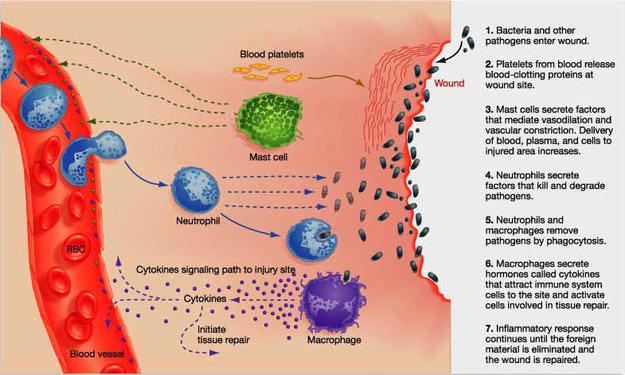
1. Tissue injury occurs which causes mast cells to secret histamine.
2. Platelets clot the blood around affected area to prevent pathogens from entering.
3. Histamine causes blood vessels to dilate increasing permeability so that phagocytes migrate to affected area.
4. Phagocytes engulf bacteria and break it down.
- An inflammatory response occurs in tissues where cells are killed or damaged by physical injury or invading pathogens. This response occurs due to mast cells detecting chemicals released from the damaged tissue. Firstly, platelets form clotting factors which seals affected area so further entry of pathogens is restricted. Mast cells secrete histamine causing nearby blood vessels dilate. As the blood vessels have become more permeable, agents such as phagocytes are able to squeeze through the membrane to the infected area, promoting an increase of fluid to the area. The phagocytes such as macrophages and neutrophil engulf the bacterium present in the infected area so that it can be broken down, resulting in the damaged tissue healing.
- The inflammatory response is carried out so that pathogens that have entered the body are broken down and digested so that they are not harmful to the body.
- As there is a greater blood supply to affected area, it caused inflamed area to become hot, swollen and red.'
Agents Involved
- Mast Cells
- Histamine
- Platelets
- Phagocytes
Stages of the Inflammatory Response
1. Tissue injury occurs which causes mast cells to secret histamine.
2. Platelets clot the blood around affected area to prevent pathogens from entering.
3. Histamine causes blood vessels to dilate increasing permeability so that phagocytes migrate to affected area.
4. Phagocytes engulf bacteria and break it down.
Interferon
- Natural proteins that are produced by the cells of the immune systems of most animals in response to challenges by foreign agent.
- Interferon is an antiviral chemical that are secreted by infected cells spreading to near by cells that take it up and begin to produce antiviral enzymes which degrades the DNA. The cells exposed to interferon prevent the nucleus from making more copies of the viral DNA and therefore prevents the virus from colonising near by cells and spreading further.
Complements
- Complements proteins are a system of proteins continually present in blood plasma.
- It assists phagocytes to recognise foreign antigens by attaching to the pathogen acting as a marker which attacks more phagocytes. Compliments are able to lyse bacterial cell walls although are useless against viruses. When the bacterial cell context spills into the host more macrophages are attracted.
